Exceptional Care,
Tailored To You
We Want You To Live Life to the Fullest
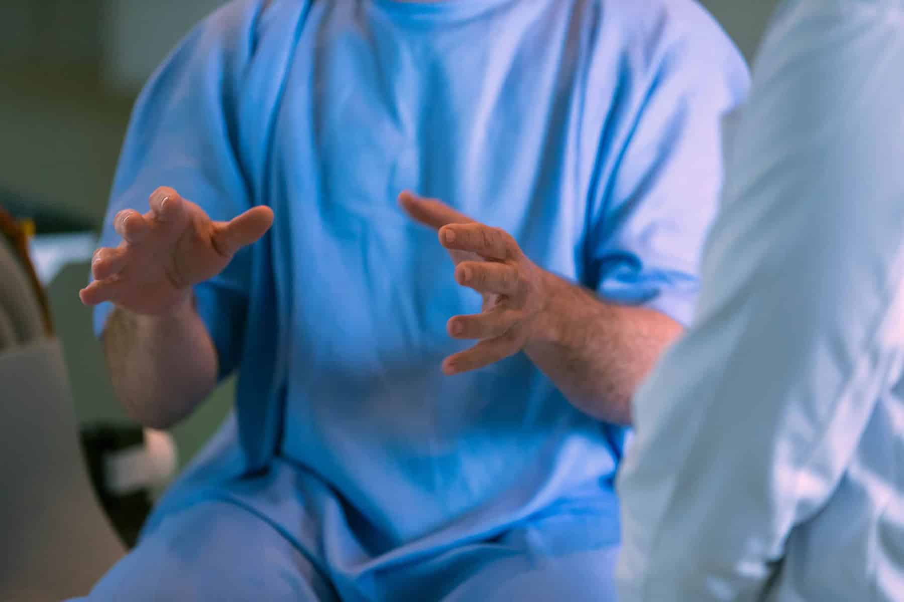
Precision’s integrated team of specialists bring innovation, experience, skill and compassion. We strive to deliver brain and body science at its best in the areas of Neurosurgery and Spinal Surgery, Neurology, Pain Management, Orthopaedic Surgery, Occupational Medicine and Rehabilitation Medicine.
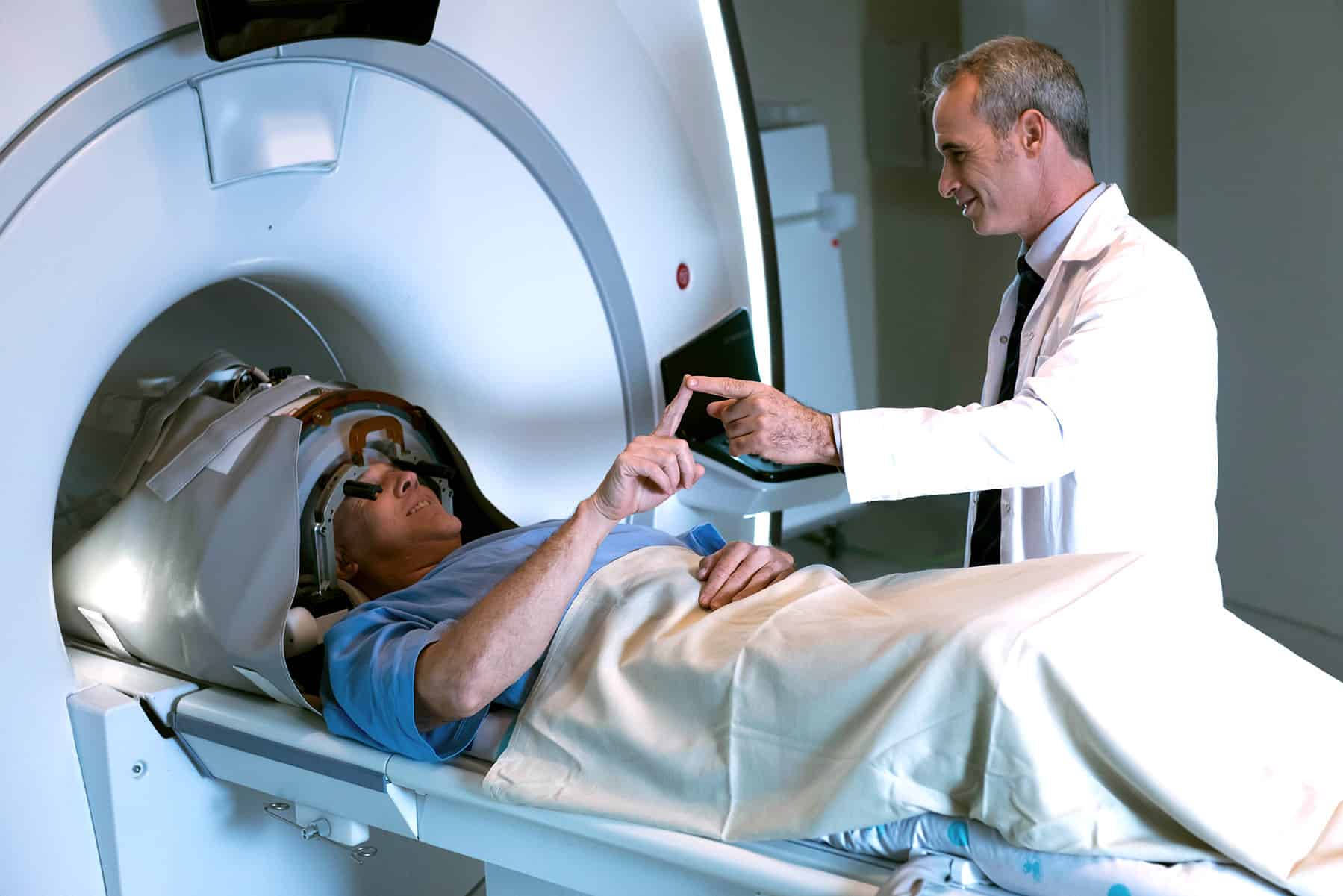
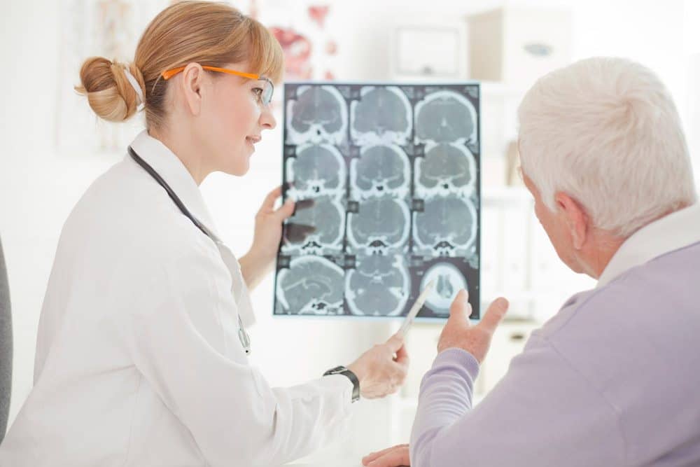
The Deep Brain Stimulation Clinic
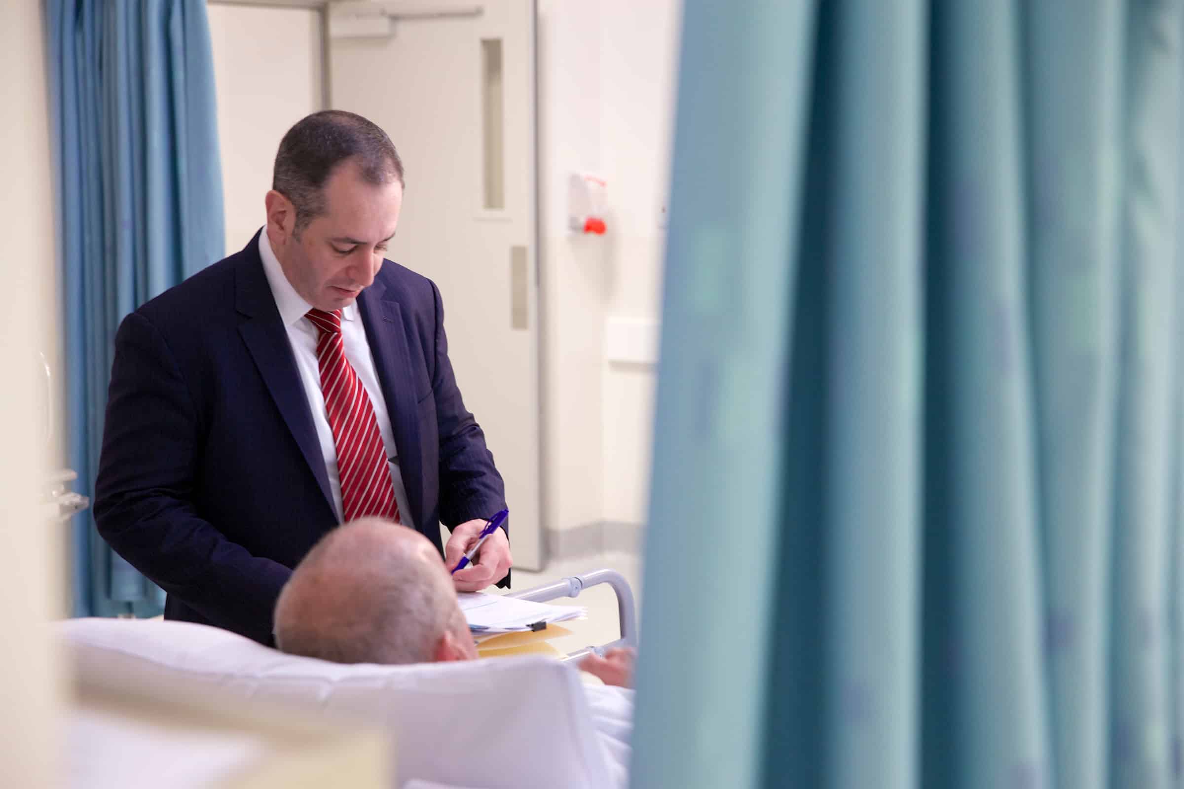
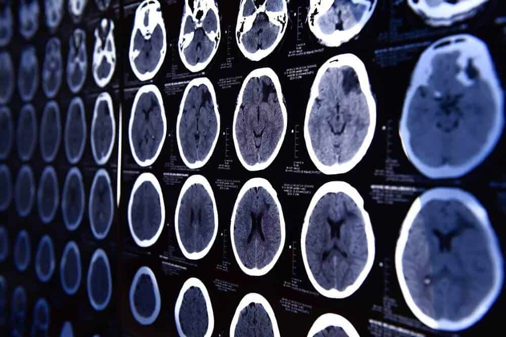
The Deep Brain Stimulation Clinic at Precision Spine & Pain Centre includes expert neurosurgeons and neurologists who perform deep brain stimulation for movement disorders and other conditions.
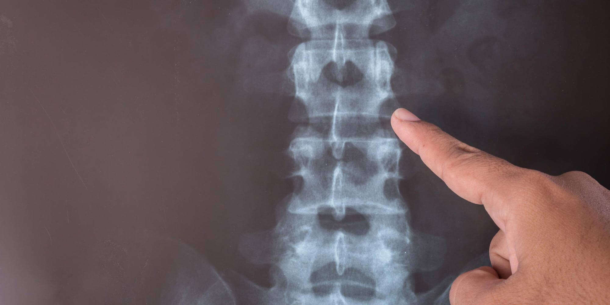
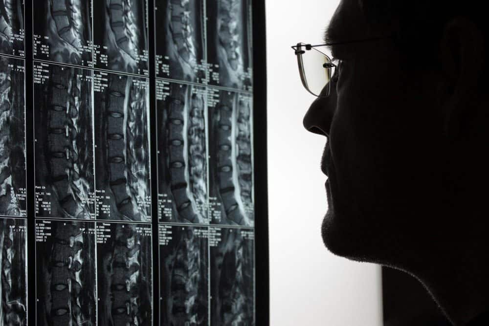
Precision Comprehensive Spine Clinic
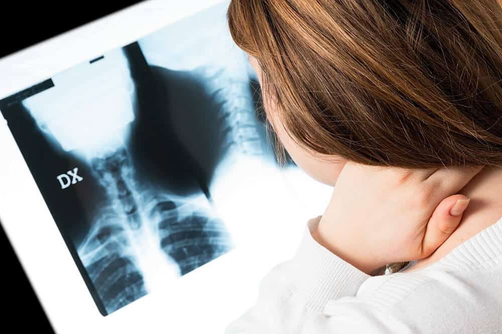
The Comprehensive Spine Clinic at Precision Brain Spine and Pain Centre is a unique, highly specialised and multidisciplinary medical clinic dedicated to the assessment and customised treatment of individuals with a range of spinal conditions.
Learn more >
Where We Are
Melbourne & Victoria
Precision Brain Spine & Pain Centre has over 20 locations around Melbourne and across Victoria. With more than 20 specialists covering a range of key specialties, we offer patients integrated care of the highest standard.
Tasmania
Precision Brain Spine and Pain Centre has consulted in Tasmania for over 10 years. Our Tasmanian hub is in Launceston, with our neurosurgeons, spinal surgeons, rehab specialists, pain specialists and neurologists providing world class treatment in several locations.
Queensland Gold Coast & Brisbane
Precision Brain Spine and Pain offers services in Brisbane, Gold Coast and Mackay. Our model of multidisciplinary healthcare includes neurosurgeons, neurologists, pain specialists, spine surgeons, and rehabilitation specialists.
Precision Brain, Spine and Pain Centre’s integrated healthcare provides more than one approach to your needs. We advocate a sophisticated strategy to deal with neurosurgical, spinal, neurological, orthopaedic and conditions requiring pain management.